Feeds:
Posts
Comments

Archive for September, 2006

brain-cover.gifThe beat goes on. The new issue of Brain (Vol. 129, No 10) contains six papers on various neurological disorders of music processing, plus a great commentary by Oliver Sacks, “The power of music”. Since I personally work on neurobiological mechanisms underlying aesthetic preference formation, I was most intrigued by a paper by Nathalie Gosselin et al., entitled “Emotional reponses to unpleasant music correlate with damage to parahippocampal cortex”. (If you don’t have a subscription to Brain you can download the paper from the Peretz Lab homepage.) Here’s the abstract:

Music is typically a pleasurable experience. But under certain circumstances, music can also be unpleasant, for example, when a young child randomly hits piano keys. Such unpleasant musical experiences have been shown to activate a network of brain structures involved in emotion, mostly located in the medial temporal lobe: the parahippocampal gyrus, amygdala, hippocampus and temporal pole. However, the differential roles of these regions remain largely unknown. In this study, pleasant and unpleasant musicwas presented to 17 patients with variable excisions of the medial temporal lobe, as well as to 19 matched controls. The pleasant music corresponded to happy and sad selections taken from the classical instrumental repertoire; the unpleasant music was the dissonant arrangement of the same selections. Only patients with substantial resections of the left or right parahippocampal cortex (PHC) gave highly abnormal judgements to dissonant music; they rated dissonant music as slightly pleasant while controls found it unpleasant. This indifference to dissonance was correlated with the remaining volume in the PHC, but was unrelated to thevolume of the surrounding structures. The impairment was specific: the same patients judged consonant music to be pleasant, and were able to judge music as happy or sad. Furthermore, this lack of responsiveness to unpleasantness was not due to a perceptual disorder, because all patients were able to detect intentional errors in the musical excerpts. Moreover, the impairment differed from that induced by amygdala damage alone. These findings are consistent with a two-dimensional model of defensive response to aversive stimuli, in which the PHC and the amygdala subserve different roles.

It is great to see that systematic neuropsychological studies of aesthetic preference formation are starting to appear, supplementing the growing number of imaging studies. How about a book on this topic, Sacks?

-Martin

Read Full Post »

chauvet.jpg

Many people are interested in the new, emerging field of neuroaesthetics – the attempt to use neuroscience to understand art and aesthetic behaviour. It is not an easy field to come to as an outsider, though. First of all, at the moment neuroaesthetics is not so much a coherent field (with textbooks and so on) as a collection of researchers with an individual interest in illuminating the neural underpinnings of art behaviour – and what these researchers take “neuroaesthetics” to mean differ rather widely. Secondly, although quite a lot has been written on neuroaesthetics in the last ten years, there is really no representative publication where newcomers can become acquainted with all the problems and research data pertinent to neuroaesthtics (for the reasons stated above).

I therefore thought that I should ease the way for the interested reader by listing a number of books that can serve as a first introduction to the world of neuroaesthetics. I have chosen to only list more or less popular books, not specialist papers, for two reasons: first, since this list is meant as an introduction, the material on it should not be too difficult; second, listing all relevant research papers is simply impossible within the framework of a short blog post. Choosing to highlight only some papers, leaving out others, would surely also make me unpopular with researchers around the world!

Neuroaesthetics can be thought of as a part of a more general study of art and aesthetics as a biological phenomenon. I will follow other proponents of this view (such as Tecumseh Fitch) in calling this broader approach bioaesthetics. The overall goal of bioaesthetics is to answer the three basic biological questions – what?, how?, why? – in regard to aesthetic behaviour in humans: what is art and aesthetics?; how does art and aesthetics spring from the brain?; and why did this cognitive ability evolve in humans? Neuroaesthetics is predominantly concerned with question number 2. In the list that follows below I will also mention a number of books that discuss the other two questions.

What is aesthetics?

Archaeological and anthropological research can help us answer such fundamental questions as when the first works of art appeared in the fossil record, what characterizes them, and who created them. It is of rather great importance to know what function(s) the first art objects had since that function reflects the cognitive capacities of those who created them. Randall White’s book from 2003, Prehistoric art (Harry N. Abrams), gives a fine overview of the when and what. Steven Mithen’s The prehistory of the mind (Thames and Hudson 1996) and David Lewis-Williams’ The mind in the cave (Thames and Hudson 2004) contain interesting speculation on the question of function and cognitive capabilities.

dancers1.jpgEqually important is ethnographic studies of what constitutes art in different contemporary societies. Much debate on “the nature” of art takes its departure from wholly theoretical considerations of what features define art. From a biological perspective it is much more interesting to know what people actually do when they create of experience art. Unfortunately, I know of no ethnographical survey, covering all the world’s cultures. However, in her books on the evolution of art Ellen Dissanayake has several great discussions of what art behaviour actually amounts to in different cultures. See especially her first two books, What is art for? (University of Washington Press 1988) and Homo Aestheticus (University of Washington Press 1992).

To these descriptions of art behaviour we should of course add the controlled investigations of experimental aesthetics. Sadly, most of the books trying to review of this psychological research tradition are rather old and outdated, but a short and idiosyncratic introduction to the field can be found in Robert Solso’s last book The psychology of art and the evolution of the conscious brain (Bradford Book 2003), which exclusive focus is on visual art, though. (Books on music are mentioned in the next section.)

How does art and aesthetics spring from the brain?

Neuroaesthetic research on how the brain gives rise to art and aesthetic behaviour can be divided up in three areas of interest: (1) Representation, (2) Emotion, and (3) Creativity.

Research on representation deals with the question of how the brain transforms perceptual inputs into mental representations – images, musical structures, etc. Since the different art forms – visual art, music, literature, dance, etc. – target different perceptual systems most researchers tend to focus on only one modality, especially vision or music. Good books on visual art are Semir Zeki’s Inner vision (Oxford University Press 1999) and Margaret Livingstone’s Vision and art (Harry N. Abrams 2002). An introduction to music research can be found in Isabelle Peretz & Roberts Zatorre (Eds.), The cognitive neuroscience of music (Oxford University Press 2003), and Daniel Livitin’s new book This is your brain on music (Dutton 2006). No books have yet been published on the cognitive neuroscience of literature – a great loss – but a few books on literature written from the point of view of cognitive science do exist, including Suzanne Nabantian’s Memory in literature (Palgrave Macmillan 2003) and Liza Zunshine’s Why we read fiction (Ohio State University Press 2006). This lack of books on literature written from the perspective of neuroscience is mostly due to the fact that, though there is a lot of neuroscientific research on language as such, almost no experiments yet have attempted to test specific literary questions. The same thing goes for dance and architecture as well (although some research appears to be forthcoming).

vermeer_painter.jpgResearch on emotion and art is a rather recent phenomenon and I know of only one book that explicitly deals with this topic, the book Music and emotion, edited by Patrick Juslin and John Sloboda (Oxford University Press 2001). I think there is reason to expect, though, that we will soon see several new books looking into it. (As Nancy Aiken reports in the comments to this post, her 1998 book, The biological origins of art, also deals with the question of how art elicits emotional responses. I am sorry to say I haven’t read that book yet, though.) In principle the field of emotion and art can be subdivided into two different problems: (1) How are emotions emulated by works of art? (2) How does the brain attach an aesthetic value to works of art? It is well known that a lot of art has human emotional life as its topic – think of romantic comedies, stories of vengeance and so on. Without the ability to induce these emotions in the viewer or reader such art works would simply be meaningless. So the ability of works of art to activate the brain’s emotional system is central to art. At the same time, art also activates the brain’s reward system, giving rise to such emotional reactions as feelings of beauty, ugliness, fascination, etc. Research on how such aesthetic emotions are computed by the brain is booming at the moment.

Finally, brain research on (artistic) creativity is still very much in its infancy. Several papers have been published recently investigating creative problem solving with fMRI and PET, but such research hasn’t really been translated into book presentations yet. The best new book on creativity and the brain is Kenneth Heilman’s Creativity and the brain (Psychology Press 2005). Readers interested in papers on artistic creativity will find several updated chapters in Colin Martindale, Paul Locher & Vladimir Petrov (Eds), Evolutionary and neurocognitive approaches to aesthetics, creativity and the arts (Baywood 2006) and Paul Locher, Colin Martindale & Leonid Dorfman (Eds), New directions in aesthetics, creativity and the arts (Baywood, in press).

I should also mention that in 2004 and 2005 Frank Clifford Rose and Dahlia Zaidel published two fascinating books collecting case stories and patient data casting further light on the issue of representation from the point of view of neuropsychology: Neurology and the arts (Imperial College Press 2004) and Neuropsychology of art (Psychology Press 2005).

Why did aesthetic cognition evolve in humans?

The evolutionary question of why aesthetic cognition evolved in humans is informed by several lines of evidence: archaeological findings, comparative studies of similarities and differences in cognitive behaviour between humans and other animals, genetics, etc. Researchers are often trying to identify either principles of sexual selection or natural selection as the driving force of the evolution of aesthetic cognition. The name most often associated with sexual selection – besides Darwin who first suggested it as a principle of evolution in The descent of man (1871) – is Geoffrey Miller who published the influential book The mating mind in 2000 (William Heinemann). The doyenne of adaptationist aesthetic studies (studies searching for natural selection forces) is Ellen Dissanayake who, apart from the two books already mentioned, published Art and intimacy in 2000 (at the University of Washington Press). The adaptationist approach has spawned quite a few publications in the last ten years, especially concerning the evolution of literature. Two good books on this topic is Joseph Carroll’s Literary darwinism (Routledge 2004) and the anthology The literary animal, edited by Jonathan Gottschall and D.S. Wilson (Northwestern University Press 2005).

chimp-painting.jpgIn addition to these books a number of publications dealing specifically with music have appeared very recently. The first, an anthology edited by Nils Wallin, Björn Merker and Steven Brown, entitled The origins of music (The MIT Press 2001) contains a wealth of different approaches, whereas Steven Mithen’s book The singing neanderthals from 2005 (Weidenfeld & Nicolson) promotes only one hypothesis.

As can be seen, the literature on bioaesthetics is rapidly growing and the probably only gain momentum in the coming years. It will be interesting to see if someone will attempt to synthesize research on all three questions, including research on all art forms, in one tome sometimes in the future.

-Martin

UPDATE. I have changed the embarassing mistitling of Mithen’s book pointed out by Geraldine in the comments. I have also fixed a couple of spelling errors.

There are clearly other relevent books out there which I haven’t mentioned. I encourage you all to suggest additional good titles in the comments section. I would personally be most interested in hearing of French and German books relevant to neuroaesthetics from readers speaking these languages.

Read Full Post »

mysticfigure.jpgA new report in Nature demonstrates that electrical stimulation of the temporoparietal junction in the brain induces a sensation of the presence of an illusory “shadowy person”. One of the hallmarks of certain forms of schizophrenia is just this phenomenon: the eery feeling of someone’s presence. Now, it has been demonstrated in a study using electrical brain activation in a person without a history of psychiatric problems.

Basically, the study was performed on a subject that was at the preoperative stage of surgery for epilepsy. A normal procedure is to anaesthesize the patient before opening the skull, and then wake the patient up before stimulating the brain. The aim of such stimulation is to find the location of epilepsy onset, as well as to stimulate the areas surrounding this region, in order to map functionally important regions (e.g. language that are important in language). Such preoperative procedures are known to lead to a better surgical sensitivity, i.e. the ability to remove all of the abnormal tissue, and a higher surgical selectivity, i.e. avoding removal of normal tissue. In this way, neurosurgeons often evoke a number of sensations and behaviours in patients, including the disruption of speech, visual phosphenes, and memory deficits.

In the present case, the neurosurgeons found that electrical stimulation lead to a feeling of the presence of another person. Moreover, the patient reported that this figure was taking the same posture as herself, and even sometimes interfering with a task she was performing:

When stimulated (…) the patient had the impression that somebody was behind her. Further stimulation induced the same experience, with the patient describing the “person” as young and of indeterminate sex, a “shadow” who did not speak or move, and whose position beneath her back was identical to her own (“He is behind me, almost at my body, but I do not feel it”). (…) Further stimulations [other location] were applied while the seated patient performed a naming (language-testing) task using a card held in her right hand: she again reported the presence of the sitting “person”, this time displaced behind her to her right and attempting to interfere with the execution of her task (“He wants to take the card”; “He doesn’t want me to read”).

Stimulation of the temporoparietal junction (shown with an arrow in the image above) thus seems to distort some kind of body image, or maybe even efference copy (PDF) of self-actions. Both functions that are dramatically affected in abnormal brain states following certain kinds of delusional schizoprenia and brain injury.

The finding also nicely relates to the Swiss group’s earlier study combining EEG, TMS and the study of an epilepsy patient, where it was found that disruption of the temporoparietal junction function led to an “impaired mental transformation of one’s own body”. Here, the researchers concluded that:

the [temporoparietal junction, TPJ] is a crucial structure for the conscious experience of the normal self, mediating spatial unity of self and body, and also suggest that impaired processing at the TPJ may lead to pathological selves such as [out-of-body experience].

Here is the full story:

Induction of an illusory shadow person
By Arzy et al
Nature 443, 287

Stimulation of a site on the brain’s left hemisphere prompts the creepy feeling that somebody is close by.

The strange sensation that somebody is nearby when no one is actually present has been described by psychiatric and neurological patients, as well as by healthy subjects, but it is not understood how the illusion is triggered by the brain1, 2. Here we describe the repeated induction of this sensation in a patient who was undergoing presurgical evaluation for epilepsy treatment, as a result of focal electrical stimulation of the left temporoparietal junction: the illusory person closely ‘shadowed’ changes in the patient’s body position and posture. These perceptions may have been due to a disturbance in the multisensory processing of body and self at the temporoparietal junction.
Top of page

1. Laboratory of Cognitive Neuroscience, École Polytechnique Fédérale de Lausanne, Lausanne 1015, Switzerland
2. Presurgical Epilepsy Evaluation Unit, University Hospital, Geneva 1211, Switzerland
3. Department of Neurology, University Hospital, Geneva 1211, Switzerland
4. Center for Cognitive Neuroscience, Dartmouth College, Dartmouth, New Hampshire 03755, USA

-Thomas

Read Full Post »

amygdala.jpgCan a brain scan reveal your relationship to your mother? According to a recent study, this may well be the case.

One of the theories in modern psychology is about the relationship between a child and her parent, especially the mother. Among such attachment theories is the original theory by John Bowlby. For a good description of attachment theory see Wikipedia. There’s also a good article (PDF) in Developmental Psychology on the history of attachment.
Basically, the theory of attachment demonstrated that the dyadic relationship between the infant and the mother can take the form of different styles — attachment styles. Such styles include secure attachment where the infant can use the mother as a secure base from which the immediate environment is explored. Insecure attachment, however, can come in different forms, including avoidant, ambivalent and disorganized (see also here). Studies have shown that the attachment style at birth is likely to influence the social functions in adulthood, and that the attachment style in one female is inherited by her offspring through a process called transmission (see also this excellent paper). Normally, this transmission is thought to be socially transmitted, although I think it’s a dubious conclusion since children are both genetically and socially related to their mother. However, a convincing study (PDF) in 2003 by Bokhorst showed that while genetic influence on temperament was relatively high, the influence on attatchment style was negligible.

But let’s get to the case: does attachment style demonstrate measurable effects on the brain? Indeed, this is what Erwin Lemche and colleagues found in a study using functional MRI. Based on previous findings that insecure attachment is related to heightened sympathetic nervous system activity (e.g. heart rate increase and cortisol secretion), Lemche et al. demonstrated that performance during a stress, relative to a neutral, prime stimulus condition involved bilateral amygdalae activation.

The subjects were shownn two series of 32 sentence statements describing self-centred or other-centred information. They had to report whether they agreed or disagreed with the statements by pressing a button. Before the presentation of the target sentences, subliminal messages with negative content were presented on some occasions (stress condition), or with nonsense sentence content (neutral condition). For example, the negative prime could be “My mom rejects me” presented for 30 milliseconds. In the neutral condition the prime could be “Ym umu jrecest em”, also presented for 30 milliseconds.

The activation of the amygdalae after negative primes was the same for all subjects. However, for those subjects who demonstrated an insecure attachment style (determined by the Adult Attachment Interview) the amygdalae activation levels was significantly higher when presented with the unconscious negative primes.

So having an insecure attachment style leads to higher activation to attachment-related primes. Taken together, this result demonstrates a role for amygdala in mediating attatchment relevant behaviour. Indeed, it is interesting to see how a phylogenetic “old” limbic structure is involved in an interpersonal psychological process, which is normally thought to involve more prefrontal cortical regions.

-Thomas

Read Full Post »

300px-us10dollarbillnew.JPGThe September 18 issue of The New Yorker contains a short introduction to neuroeconomics. It is a nice enough article without to many obvious errors (the fact that the author attributes the striatum to the limbic system instead of to the basal ganglia is probably only of concequence to anatomists!). It tells the now familiar story of how the classical view of homo economicus has been challenged by the experimental work of Kahneman, Tversky and other members of the so-called behavioural economics field. It then goes through some of the recent imaging experiments on risk aversion, punishment of unfair collaborators, and trust, with quotes from several of the key neuroeconomics players: Colin Camerer, George Loewenstein, David Laibson, Jonathan Cohen, Ernst Fehr, and Paul Glimcher.

To people already acquainted with the field the most interesting parts of the article is probably a section where Laibson and Cohen ponders possible policy implications of neuroeconomics research, and a section where Cohen and Glimcher are reported to nurture opposing views of how “emotion” and “cognition” interacts in decision-making situations. Briefly, Cohen views emotion and cognition as two separate systems that may at times “compete” for control, whereas Glimcher disputes such a clear-cut separation. Personally, I would have loved to hear more about this issue.

Here are two links to some earlier posts on neuroeconomics:

Risk aversion is rewarding!

Neurofinance

-Martin

Read Full Post »

einstein.jpgWhat characterizes Albert Einstein‘s brain? Why did he become a genius? Can we trace it down to brain-related factors? A growing literature on the relationship between intelligence and brain structure and function has demonstrated several relationships. Those studies, however, are typically based on comparison of brains of high versus mean IQ groups. Studying individual geniuses and what is special about their brains are rare in the scientific literature. However, there are a few exceptions.

In a study by Colombo et al in Brain Research Reviews, the brain of Albert Einstein is studied and compared to four age-matched individuals without any known neurological or psychiatric symptoms. The researchers found that

Einstein’s astrocytic processes showed larger sizes and higher numbers of interlaminar terminal masses, reaching sizes of 15 μm in diameter.

And they further notice that

These bulbous endings are of unknown significance and they have been described occurring in Alzheimer’s disease

…which would mean that, if anything, the size and number of interlaminal terminal masses in Einstein’s brain would make it more like an Alzheimer-patient than like a genius.

Colombo and colleagues are indeed sceptic about the findings and interpretations in the literature on Einstein’s brain. But why do this study in the first place? I’m baffled — to put it mildly — that this kind of study is published in a well-esteemed (well, any) scientific journal. This due to especially three factors relating to the validity of the study:

  1. There are only four control subjects. This provides no information about what the population as a whole looks like for the given brain measurement. IOW, we cannot know anything about the natural variance in the population of our measures, let alone know much about the mean value. Given this, the up-to-15-μm-diameter interlaminal terminal masses means nothing, since we cannot know anything about whether Einstein’s brain is special
  2. One is studying an old and degenerated brain. The fact that the study is of Einstein’s brain at a high age (76 years) seems irrelevant to discover what made his brain so special during the age at which he formulated and developed his theoretical ideas, i.e. decades earlier. This period not only includes the time at which he wrote about relativity, but also an earlier and less known period when he wrote the Annus Mirabilis Papers. This latter series of articles are concerned with the photoelectric effect, also recognized as papers that alone deserve a Nobel prize. And those papers were even written by Einstein during his spare time!
  3. Why not study a group of geniuses? Indeed, why only rely on one data point? Why not include a larger sample of geniuses, not only from physics, but from other sciences? Einstein was not the only genius around. Living in Denmark and passing through the Copenhagen University physics buildings regularly, I am immediately reminded of Niels Bohr, a contemporary to Einstein that matches Einstein’s genius in every respect. It would thus be much more interesting to see a group study of geniuses. It might be hard to do a genius post-morten study for both practical and ethical reasons, but one can do in vivo studies of today’s geniuses, right? One data point, even if it’s Einstein, is really not enough. Doing a group study we could also ask questions such as whether there are differences between male and female geniuses, or whether the develop and age different from the general population.

Einstein’s brain is indeed an interesting topic, but in order to make valid inferences from a study of his brain, we should consider including his brain among a number of related geniuses. Doing any kind of study of one genius’ brain is unlikely to produce any valid finding at all.

-Thomas

Read Full Post »

A conscious veggie?

lockedin.jpgIt should be mentioned that there is a highly controversial paper out in this week’s Science. Here, a study by Dr Adrian Owen shows that a patient that meets the criteria for vegetative state shows what can be thought of as signs of conscious life. You can see the MindHacks coverage of this story here. basically, the researchers asked their patient to imagine playing tennis or moving around her home and found that:

the patient activated predicted cortical areas in a manner indistinguishable from that of healthy volunteers

The researchers take this as a sign of the patient being conscious while still being unable to communicate, let alone perform any volutary behaviour. In this sense, it seems that the patient rather meets the criteria for locked-in syndrome. The study thus raises the question of whether this patient — indeed, any PVS patient — is conscious, and whether the diagnostic criteria are yet poorly defined and the symptoms similarly poorly understood.

Nevertheless, it should be considered an open question whether we should accept this finding as a true sign of consciousness in the patient. This is also covered nicely in a comment by Naccache in the same issue of Science. Pointing out some of the shortcomings of this study, as well as contrasting it to the many studies showing specific changes in PVS contrasted to normal consciousness, Naccache concludes:

Though not totally convincing on the issue of consciousness, the Owen et al. work paves the way for future functional brain-imaging studies on comatose and vegetative state patients. One can imagine probing each of the psychological properties of conscious processing listed above, and even trying to collect subjective reports by modifying the experimental paradigm.

As I have written earlier, this is indeed the case: we do not have a clear concept of the distinctions between coma, vegetative states, minimally conscious states or locked-in syndrome. Only during the past few years we have seen a dramatic increase in our understanding of these disorders. It is no doubt that our understanding will increase in the same manner during the next many years.

I have asked Dr. Owen and his co-author, Steven Laureys about this finding, and hope to get back to you with their replies shortly. In the meantime, you might be interested in this article (PDF) that Laureys and I co-authored with prof. Bernard Baars in TINS.

UPDATE: Nature has a podcast interview with Dr. Owen here.

-Thomas

Read Full Post »

In an interesting paper in the latest version of Progress in Neurobiology, Yuri I. Arshavsky from UCSD writes about the epistemological dualism that exists in modern neuroscience. basically, Arshavsky claims that there is a covert dualism in the way that neuroscientists are treating mind-related topics, especially the study of “consciousness”. Indeed, as he claims:

This covert dualism seems to be rooted in the main paradigm of neuroscience that suggests that cognitive functions, such as language production and comprehension, face recognition, declarative memory, emotions, etc., are performed by neural networks consisting of simple elements.

This might initially sound a bit strange. Is not cognitive functions such as face perception due to operational simple elements? Face perception as such is a combination of many simple processes that operate in unison. So what is Arshavsky proposing? Indeed he suggests the existence of a certain kind of brain cells:

(The) performance of cognitive functions is based on complex cooperative activity of “complex” neurons that are carriers of “elementary cognition.” The uniqueness of human cognitive functions, which has a genetic basis, is determined by the specificity of genes expressed by these “complex” neurons. The main goal of the review is to show that the identification of the genes implicated in cognitive functions and the understanding of a functional role of their products is a possible way to overcome covert dualism in neuroscience.

So there should exist a subset of neurons that integrate information from a variety of input. This sounds strange, since all neurons integrate inputs from thousands of inputs, many from a large variety of inputs. So what are complex neurons? Here, we are told that:

(…) neural networks involved in performing cognitive functions are formed not by simple neurons whose function is limited to the generation of electrical potentials and transmission of signals to other neurons, but by complex neurons that can be regarded as carriers of “elementary” cognition. The performance of cognitive functions is based on the cooperative activity of this type of complex neurons.

In this way, complex neurons seem to be integrative neurons, i.e. cells that integrate information from a variety of processes. This could include the multi-modal neurons found in the functional sub-structures of the medial temporal lobe, such as the hippocampus, perirhinal, entorhinal and temporopolar cortex. But would it not mean the colour processing nodes in the visual cortex? Which IMO leads us back to a basic question: what is a functional unit in the brain. yes, the neuron is a basic building block of information processing in the brain. But what is special about language, memory and so forth in the brain?

It is possible that Arshavsky is not radical enough: what we should seek out is to avoid using generalistic and folk-psychological concepts in the first place. We should possibly not study “language”, “memory” or “consciousness”, since these concepts will always allude to fundamental assumptions of “language-ness”, “memory-ness” and “consciousness-ness”, IOW that there is something more to explain after we have found out how the brain produces what we recognize and label a cognitive function.

Maybe neuroscientists are not using a poor strategy after all? Maybe ignoring the past history of philosophy of mind is the best solution. I’m not sure (nor am I sure that I represent Arshavsky’s view properly). But how we choose to label a cognitive function depend on our past historical influence and learning, as well as our current approach.

-Thomas

Read Full Post »

Here is a great story: human imitation has been known to be present in newborns, supporting a notion of the human race being predisposed to social interaction. However, an obvious question of whether this is also the case in non-human primates below our closest evolutionary relatives has not been asked. Until now. In an excellent study by Pier Ferrari and colleagues in PLoS Biology, imitation of facial expression is demonstrated in neonatal monkeys (that disappeared after approx. 7 days).

From ScienceDaily we can read:

Ferrari et al. tested 21 baby rhesus monkeys’ response to various experimental conditions at different ages (one, three, seven, and 14 days old). Infants were held in front of a researcher who began with a passive expression (the baseline condition) and then made one of several gestures, including tongue protrusion, mouth opening, lip smacking, and hand opening.

Day-old infants rarely displayed mouth opening behavior, but smacked their lips frequently. When experimenters performed the mouth opening gesture, infants responded with increased lip smacking but did not increase any other behavior. None of the other stimuli produced significant responses. But by day 3, matched behaviors emerged: infants stuck out their tongues far more often in response to researchers’ tongue protrusions compared with control conditions, and smacked their lips far more often while watching researchers smacking theirs. By day 7, the monkeys tended to decrease lip smacking when humans performed the gesture, and by two weeks, all imitative behavior stopped.

Here is an example from the article:

And from the abstract:

Our findings provide a quantitative description of neonatal imitation in a nonhuman primate species and suggest that these imitative capacities, contrary to what was previously thought, are not unique to the ape and human lineage. We suggest that their evolutionary origins may be traced to affiliative gestures with communicative functions.

UPDATE: Jown Hawkes has an in-depth presentation and discussion of this study.

-Thomas

Read Full Post »

brainsurgery.jpg
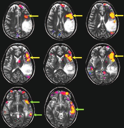
Brain tumors are a huge problem in neurosurgery. Not only do you have to take into consideration the delicate network of blood supply to the brain that can ultimately lead to further damage to the brain. In addition, the tumor is placed with in a meshwork of cognitive functions. Cutting too much on one side of the tumor can lead to amnesia, too much of another part can lead to aphasia.

To alleviate this problem, MRI is currently being used to identify the tumor through conventional structural scans. In addition functional MRI can be used to identify vital functional nodes that borders to the tumor. In this way, neurosurgery can use a better estimate of the tumor’s position and extent, as well as avoid functional centres. In all, the precision of neurosurgery has improved dramatically with the use of MRI.

In an article in Radiology, a study now shows that the use of structural and functional MRI in preoperative surgical planning both leads to more precise and more efficient surgery. As a brief resume in Medscape.com reports:

In six cases, the neurosurgeon reported that functional MRI results led to a more complete resection, whereas two patients required a smaller craniotomy than had been planned. The surgeons also noted that surgical time was reduced by 15 to 60 minutes in 22 patients. Invasive imaging that would have been required for four patients was avoided.

In practice this has a tremendous impact for the livesof the patients. With the use of preoperative MRI brain tumors can now be more fully ablated, and at the same time patients will have a lower chance of suffering unwanted dysfunctions. From the Radiology paper, we can see this in one female patients. From the description of the patient:

preoperativefmri.jpg

Recurrent left parietal lobe anaplastic astrocytoma in 37-year-old right-handed woman. Surgery was not initially planned because of presumed involvement of receptive speech area. Left inferior and middle frontal gyral activation (yellow arrows) is consistent with dominant expressive speech area and is located at anterior border of more cephalad component of lesion. Left superior and middle temporal gyral activation (green arrows) is consistent with dominant receptive speech area and abuts inferior border of temporal component of lesion, with superior temporal gyral activation component lying anteroinferior to lesion. Biopsy was performed, and no postoperative neurologic deficits were documented.

-Thomas

Read Full Post »

Older Posts »